


DNA Isolation Kit for Mammalian Blood
DNA Isolation Kit for Mammalian Blood; Instructions For Use
Isolation of DNA from whole blood can be difficult because blood is a complex mixture containing cells, proteins, metabolites, etc. Most of the cells (>99%) are erythrocytes (red blood cells) which lack nuclei, and therefore, possess no DNA. Leukocytes
(white blood cells) contain nuclei and DNA. Therefore, DNA from blood must be isolated from one of three types of leukocytes: monocytes, lymphocytes, or granulocytes.
Sample material:
Human whole blood (treated with sodium heparin,
EDTA or sodium citrate): 1 to 10 mL
Isolated lymphocytes: 1 to 10 mL
Isolated buffy coat: buffy coat from 10 to 20 mL whole blood, containing at least 1.0 x 10⁷ leukocytes.
Purity of isolated DNA: Average A 260 / A 280 ratio: 1.7
- 1.9
The DNA Isolation Kit for Mammalian Blood rapidly isolates DNA from 1 to 10 ml mammalian whole blood, lymphocytes, or buffy coat samples.
DNA Isolation Kit for Mammalian Blood has been used in isolation of genomic DNA from blood of asthmatic patients for single nucleotide polymorphisms genotyping.[1]
The DNA Isolation Kit for Mammalian Blood purifies DNA that is suitable for most molecular biology applications:
• PCR/long PCR
• Sequencing
• Restriction digestion
• Southern blotting
• Cloning
• Process multiple samples simultaneously.
DNA is easily purified in <90 minutes (plus 30 to 60 minutes resuspension time).
• Use a cost-effective kit.
Achieve faster DNA purification for the cost of most homebrew
methods.
• Obtain consistent and reliable results.
Analyze different sample volumes (1 to 10 mL) with varying amounts of leukocytes.
• Improve safety.
Eliminate the use of chaotropic salts, hazardous organic solvents,
and column purification steps.
• Red Blood Cell Lysis Buffer
• White Blood Cell Lysis Buffer
• Protein Precipitation Solution
Each lot of kits is function tested for the ability to purify DNA from human whole blood, followed by specific amplification of a 4.8 kb tPA fragment via PCR using the Expand Long Template PCR System. The 4.8 kb tPA product is visualized by electrophoresis on an agarose gel and compared to appropriate positive and negative controls. A single 4.8 kb tPA band of equivalent intensity to the positive control is visible. The kit buffers are also tested for the absence of DNase contamination. Each solution is incubated with 1 μg pBR322 DNA for 6 hours at +37°C. The DNA is then visualized by electrophoresis on an agarose gel, and then compared to a positive control to determine if any linear or nicked plasmid DNA is visible.
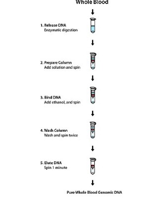
The DNA Isolation Kit for Mammalian Blood procedure relies on the separation of white blood cells from whole blood via a preferential red blood cell lysis. In the presence of a strong anionic detergent, the white blood cells are lysed, and then the proteins are removed by dehydration and precipitation. The purified DNA is subsequently recovered via ethanol precipitation.
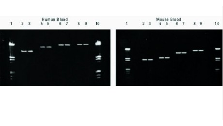
Figure 1: Amplification of large DNA fragments from mammalian blood prepared with the DNA Isolation Kit for Mammalian Blood. DNA was prepared from two research samples, one containing 10 ml human blood, the other containing 10 ml mouse blood.
Aliquots of each DNA preparation were used as templates for the amplification of several gene fragments with the Expand Long Template PCR System. PCR products were analyzed by gel electrophoresis.
Left panel: Gene fragments amplified from human
samples
Lanes 2, 3: tPA fragment (9.3 kb) amplified from 25 ng DNA
Lanes 4, 5: tPA fragment (15 kb) amplified from 50 ng DNA
Lanes 6, 7: β-globin fragment (23 kb) amplified from 100 ng DNA
Lanes 8, 9: β-globin fragment (28 kb) amplified
from 200 ng DNA
Lanes 1, 10: DNA Molecular Weight Marker III
Right panel: Gene fragments amplified from mouse samples
Lanes 2, 3: IL-2 gene (4.2 kb) amplified from 330 ng DNA
Lanes 4, 5: α-2 collagen fragment (5.6 kb) amplified from 100
ng DNA
Lanes 6, 7: α-2 collagen fragment (10.4 kb) amplified from 50 ng DNA
Lanes 8, 9: α-2 collagen fragment (15.4 kb) amplified from 100 ng DNA
Lanes 1, 10: DNA Molecular Weight Marker III
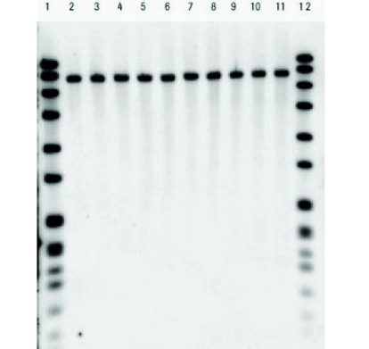
Figure 2: Southern blot analysis of DNA from various human blood samples prepared with the DNA Isolation Kit for Mammalian Blood. DNA was prepared from several research samples, including human blood containing different anticoagulants and
lymphocyte and buffy coat preparations. Ten μg of each preparation was digested with Eco RI, separated by gel electrophoresis, and transferred to a nylon membrane by Southern blotting. The n-ras gene in each sample was detected with a DIG-labeled
n-ras probe.
Lanes 2, 3: Blood sample containing sodium citrate
Lanes 4, 5: Blood sample containing heparin
Lanes 6, 7: Blood sample containing sodium EDTA
Lanes 8, 9: Buffy coat preparation
Lanes 10, 11: Lymphocyte preparation
Lanes
1, 12: DNA Molecular Weight Marker VII
Result: Each lane contained only a single hybridized band of the expected size.
For life science research only. Not for use in diagnostic procedures.
01.jpg)